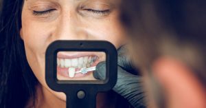
Ever wondered why some people seem to have a few extra teeth crowding their smile? Hyperdontia, the condition of having supernumerary teeth, can turn a dazzling grin into a dental dilemma. If you’re dealing with this quirky condition, you’re not alone, and the good news is that hyperdontia removal can bring your pearly whites back to their intended glory.
I’ve seen firsthand how those extra teeth can complicate everything from chewing to confidence. But fear not, because modern dental practices offer effective solutions to this peculiar problem. Ready to learn how to reclaim your smile and say goodbye to those unwelcome guests in your mouth? Let’s dive into the fascinating world of hyperdontia removal.
Key Takeaways
- Hyperdontia Overview: Hyperdontia is a condition characterized by extra teeth, known as supernumerary teeth, which can cause dental complications like misalignment, crowding, and difficulty in chewing and speaking.
- Causes: The primary causes of hyperdontia include genetic factors, such as a family history or conditions like cleidocranial dysplasia, as well as environmental factors like nutritional deficiencies, substance exposure during pregnancy, and mouth trauma.
- Types of Supernumerary Teeth: Hyperdontia can present in different forms including supplemental (extra teeth resembling normal teeth), tuberculate (barrel-shaped), conical (peg-like), and compound odontoma (clusters of tooth-like structures).
- Diagnosis Methods: Detecting hyperdontia involves both clinical examination and radiographic imaging like panoramic X-rays and cone beam computed tomography (CBCT) for accurate identification of extra teeth.
- Removal Procedures: Treatment of hyperdontia generally involves removing the extra teeth through simple extraction if they are fully erupted, or surgical extraction for impacted teeth.
- Recovery and Aftercare: Post-removal care is crucial, focusing on pain management with medications and ice packs, maintaining good oral hygiene, and attending follow-up appointments to ensure proper healing. Potential complications include infection and, in rare cases, nerve damage.
Understanding Hyperdontia
Hyperdontia refers to a dental condition where extra teeth grow in the mouth. These additional teeth, commonly called supernumerary teeth, can appear in any area of the dental arch. While many cases of hyperdontia present with just one or two extra teeth, instances with several supernumerary teeth happen as well.
These extra teeth can lead to various dental problems. Misaligned teeth are a common issue caused by hyperdontia. Individuals with the condition may also experience difficulties with chewing and speaking. In addition, crowding in the mouth can occur, leading to further complications.
Children and adults can both be affected by hyperdontia. It often becomes noticeable once the primary teeth have erupted. However, in some cases, it may not become apparent until the dentist performs an X-ray. Recognizing and diagnosing hyperdontia early helps in planning appropriate treatment.
Causes of hyperdontia include genetic factors. There is often a family history of the condition. Sometimes hyperdontia aligns with other medical conditions like cleidocranial dysplasia. If hyperdontia is suspected, consulting a dentist is crucial for an accurate diagnosis and effective treatment plan.
Treating hyperdontia usually involves removing the extra teeth. Dentists use various methods to do so. Extraction is the most common approach. Advanced dental tools and techniques ensure safe and effective removal of supernumerary teeth. Regular dental check-ups aid in monitoring and managing hyperdontia effectively.
Causes Of Hyperdontia

Hyperdontia occurs from various influences. Genetic predisposition and environmental aspects play significant roles in its development.
Genetic Factors
Genes greatly influence hyperdontia. Conditions like cleidocranial dysplasia are often tied to this dental anomaly. People with this condition may experience delayed tooth eruption and irregular bone development, leading to the formation of supernumerary teeth.
Some families exhibit a hereditary pattern where several members face hyperdontia. When extra teeth appear in parents, their children have a higher chance of developing the same issue. This suggests a strong genetic connection.
Mutations in specific genes are also critical. Research has identified that mutations in genes such as RUNX2 or AXIN2 can disrupt normal tooth development, causing additional teeth to form. It’s essential to understand these factors for accurate diagnosis and treatment.
Environmental Factors
Environmental factors also influence hyperdontia. During pregnancy, exposure to certain substances might affect tooth development. For example, excessive consumption of alcohol or illicit drugs can disrupt the normal formation of teeth in the fetus.
Childhood trauma can lead to the development of extra teeth. A direct injury to the mouth might stimulate the growth of excess teeth as the body’s way of compensating for the damage.
Nutritional deficiencies during early childhood play a role too. Lack of essential vitamins and minerals, such as Vitamin D and calcium, can impair normal dental development and result in supernumerary teeth. Ensuring a balanced diet helps mitigate this risk.
Establishing a clear understanding of these factors allows for better preventive measures and tailored treatments. Identifying the root cause aids in managing hyperdontia effectively.
Types Of Hyperdontia

Hyperdontia can manifest in various forms depending on the characteristics of the extra teeth. Understanding these types aids in accurate diagnosis and treatment plans.
Supplemental
Supplemental teeth mimic the shape and form of normal teeth. These extra teeth often appear behind the permanent teeth, making them harder to detect without X-rays. For instance, a supplemental incisor may look just like the standard central or lateral incisor, only appearing in addition to the regular set. Dentists pay special attention during check-ups to spot these teeth as they can contribute to misalignment and crowding if untreated. Removing supplemental teeth usually requires careful planning to minimize impact on the rest of the teeth.
Tuberculate
Tuberculate teeth have a barrel-like shape and sometimes contain multiple cusps. They are typically found in pairs. They usually don’t erupt into the mouth, lying instead within the bone structure. This type often impacts new tooth eruption, causing delays or eruptions in abnormal places. Identifying tuberculate teeth involves advanced imaging techniques, which pinpoint their exact location and structure. Dentists consider surgical options in such cases to prevent future complications and retain proper dental harmony.
Conical
The conical form is easily recognized due to its peg-like shape. Commonly located in the anterior region of the upper jaw, these teeth can cause neighboring teeth to crowd or become displaced. Unlike other types, conical teeth may erupt fully and become visible. This visibility makes early diagnosis simpler. Quick action is critical to ensure these teeth don’t affect the aesthetics and function of one’s smile. Orthodontists often collaborate with dentists to devise comprehensive treatment plans targeting these specific problems.
Compound Odontoma
Compound odontomas consist of multiple small tooth-like structures. These composites generally don’t resemble normal teeth but instead form clusters. Often situated in the anterior maxillary region, they can obstruct normal tooth eruption and lead to complex dental issues. Comprehensive imaging helps detect compound odontomas early on. Addressing them early often involves a combination of surgical removal and orthodontic correction to preserve overall oral health. Removing these intricate structures necessitates precision to avoid damaging adjacent teeth and bones.
Diagnosis Of Hyperdontia

Detecting hyperdontia early allows for effective management of the condition. Both clinical examination and radiographic imaging play pivotal roles in diagnosing this dental anomaly.
Clinical Examination
Dentists assess the overall oral cavity to look for abnormalities. They inspect each quadrant of the mouth for extra teeth, which may appear similar to natural teeth or have distinct shapes like barrel-like or peg-like forms. During the examination, dentists also evaluate the alignment of teeth and gum health since crowding and misalignment often accompany hyperdontia.
Sometimes, dentists utilize probes to feel for unerupted supernumerary teeth that haven’t broken through the gum surface yet. I always ensure I have a full picture of the patient’s oral health before recommending further diagnostic steps. Clinical examination alone sometimes cannot confirm the presence and exact position of all extra teeth, making further diagnostic tools necessary.
Radiographic Imaging
Dentists use advanced imaging techniques to get a comprehensive view of the mouth’s internal structure. Panoramic X-rays often provide an initial overview, highlighting any extra teeth that might not be visible during a clinical examination. Cone beam computed tomography (CBCT) offers a more detailed, 3D view, crucial for identifying the exact location and orientation of supernumerary teeth. This technology surpasses traditional X-rays in precisely visualizing teeth positioned within the jawbone.
In some cases, dentists recommend a periapical radiograph to focus on a specific area of concern. This detailed image helps in planning surgical removal if needed. By combining clinical examination and radiographic imaging, dental professionals accurately diagnose hyperdontia, ensuring personalized treatment plans for the best outcomes.
Hyperdontia Removal Procedures
Hyperdontia removal procedures depend on the location, type, and complexity of the extra teeth. Two main methods are commonly employed: simple extraction and surgical extraction.
Simple Extraction
Simple extraction suits cases where extra teeth are fully erupted and easily accessible. Dentists use local anesthesia to numb the area, ensuring a painless procedure. After numbing, they use an elevator to loosen the tooth. Once loosened, forceps remove the tooth with minimal discomfort. Recovery is generally quick, usually taking about a week. Patients are advised to follow post-extraction care instructions to avoid complications like dry socket.
If the extra teeth are small and not deeply rooted, simple extraction becomes a straightforward process. Most children with hyperdontia undergo simple extraction to prevent misalignment of permanent teeth. Not all hyperdontia cases qualify for simple extraction, especially if extra teeth are impacted or fused to the bone.
Surgical Extraction
When extra teeth are impacted or not fully erupted, surgical extraction becomes necessary. Oral surgeons perform this procedure under local anesthesia, occasionally using sedation for more complex cases. The surgical approach involves making an incision in the gum to access and remove the tooth. Bone removal around the tooth might be required if it’s deeply embedded.
Patients might experience swelling and discomfort post-surgery, but pain is manageable with prescribed medications. The healing process can take up to two weeks. It’s vital to maintain good oral hygiene during recovery to prevent infections. For patients with multiple or complex supernumerary teeth, surgical extraction ensures comprehensive removal, preserving overall oral health.
Recovery And Aftercare
Hyperdontia removal isn’t the end of the journey; it’s crucial to focus on recovery and aftercare to ensure successful results.
Pain Management
Pain management is a priority post hyperdontia removal. Over-the-counter pain relievers like ibuprofen or acetaminophen help alleviate discomfort. I recommend taking medication as prescribed by your dentist. Ice packs applied to the cheek near the extraction site can reduce swelling and numb pain. Avoid consuming hot or hard foods that might aggravate the area. If discomfort persists beyond a few days or worsens, consulting your dentist is essential.
Oral Hygiene
Maintaining oral hygiene post-surgery is critical. Start with gentle rinsing using a saltwater solution after 24 hours to keep the area clean. Continue brushing your teeth but be cautious around the extraction site to avoid irritation. Refrain from using mouthwash with alcohol, as it can irritate the healing tissues. Stick to soft foods like yogurt or mashed potatoes to avoid putting pressure on the extraction site.
Follow-Up Appointments
Follow-up appointments are necessary to monitor healing progress. Schedule a visit with your dentist within a week after the procedure. They will check for signs of infection or other complications. I find it helpful to discuss any concerns or unexpected symptoms during these visits. Regular follow-ups ensure that the extraction site heals properly, and long-term oral health is maintained.
Potential Complications
Hyperdontia removal can have several potential complications. Patients should be aware of these risks and consult their dentists for proper management.
Infection
Infections can occur after any dental procedure, including hyperdontia removal. Symptoms to watch for include swelling, severe pain, and a persistent bad taste. Keeping the mouth clean, especially after meals, reduces this risk. Dentists might prescribe antibiotics to prevent infections. I recommend following the prescribed dosage and completing the full course. Regular check-ups will help detect any early signs of infection. Saltwater rinses also aid in keeping the surgical site clean. If you notice unusual symptoms, contact your dentist immediately.
Nerve Damage
Nerve damage is another potential risk during hyperdontia removal. Patients might experience numbness or tingling in the lips, tongue, or chin. This usually subsides within a few weeks but can sometimes be permanent. Dentists carefully evaluate the position of extra teeth relative to the nerves to minimize this risk. I suggest discussing any concerns about nerve damage with your dentist before surgery. Follow their advice closely and report any prolonged numbness. Early intervention can help manage nerve-related issues effectively.
Conclusion
Hyperdontia removal is a complex but manageable process when approached with the right knowledge and care. Regular dental check-ups and early diagnosis play a critical role in effective treatment. It’s essential to follow your dentist’s recommendations for post-removal care to ensure a smooth recovery and avoid complications like infections or nerve damage. Always maintain good oral hygiene and keep an open line of communication with your dental care provider. With proper care and attention, you can navigate the challenges of hyperdontia and maintain a healthy smile.
Frequently Asked Questions
What is hyperdontia?
Hyperdontia is a condition characterized by having extra teeth beyond the normal set of 32 adult teeth. These extra teeth are called supernumerary teeth and can occur anywhere in the dental arch.
What causes hyperdontia?
Hyperdontia is generally caused by genetic factors. It tends to run in families, so if one or both parents have extra teeth, their children may also develop this condition.
How is hyperdontia diagnosed?
Hyperdontia is diagnosed through dental exams and imaging techniques such as X-rays. Dentists and orthodontists can identify the presence and position of extra teeth during these examinations.
What are the treatment options for hyperdontia?
Treatment usually involves the removal of the extra teeth. A dentist or orthodontist will create a treatment plan, which may include surgical extraction and potential orthodontic treatment to correct alignment issues.
Is removal of extra teeth necessary?
Not always. The necessity of removing extra teeth depends on their location, potential impact on dental health, and whether they cause functional or aesthetic issues. Your dentist will make a recommendation based on your specific case.
What are potential complications of hyperdontia removal?
Potential complications can include infections, nerve damage, and prolonged healing times. Dentists take precautions to minimize these risks, but it’s important to discuss any concerns prior to surgery.
How can complications be prevented post-surgery?
Maintaining oral hygiene, following prescribed antibiotic regimens, and keeping all follow-up appointments can help prevent complications. Discuss any concerns about nerve damage or other issues with your dentist beforehand.
How important are regular dental check-ups for managing hyperdontia?
Regular dental check-ups are crucial for timely diagnosis and management of hyperdontia. Early detection allows for better treatment planning and reduces the risk of subsequent complications.
What does post-removal care involve?
Post-removal care includes pain management, maintaining oral cleanliness, and attending follow-up appointments to monitor healing. Your dentist will provide specific instructions tailored to your situation.
Why is early intervention important for hyperdontia?
Early intervention helps manage hyperdontia more effectively, reducing the risk of complications such as misalignment of other teeth and jaw problems. This allows for a more straightforward and less invasive treatment process.